What Happens During a 30-Second Face Scan?
Find out what happens during a 30-second face scan that reads your vital signs through your phone camera using rPPG technology.
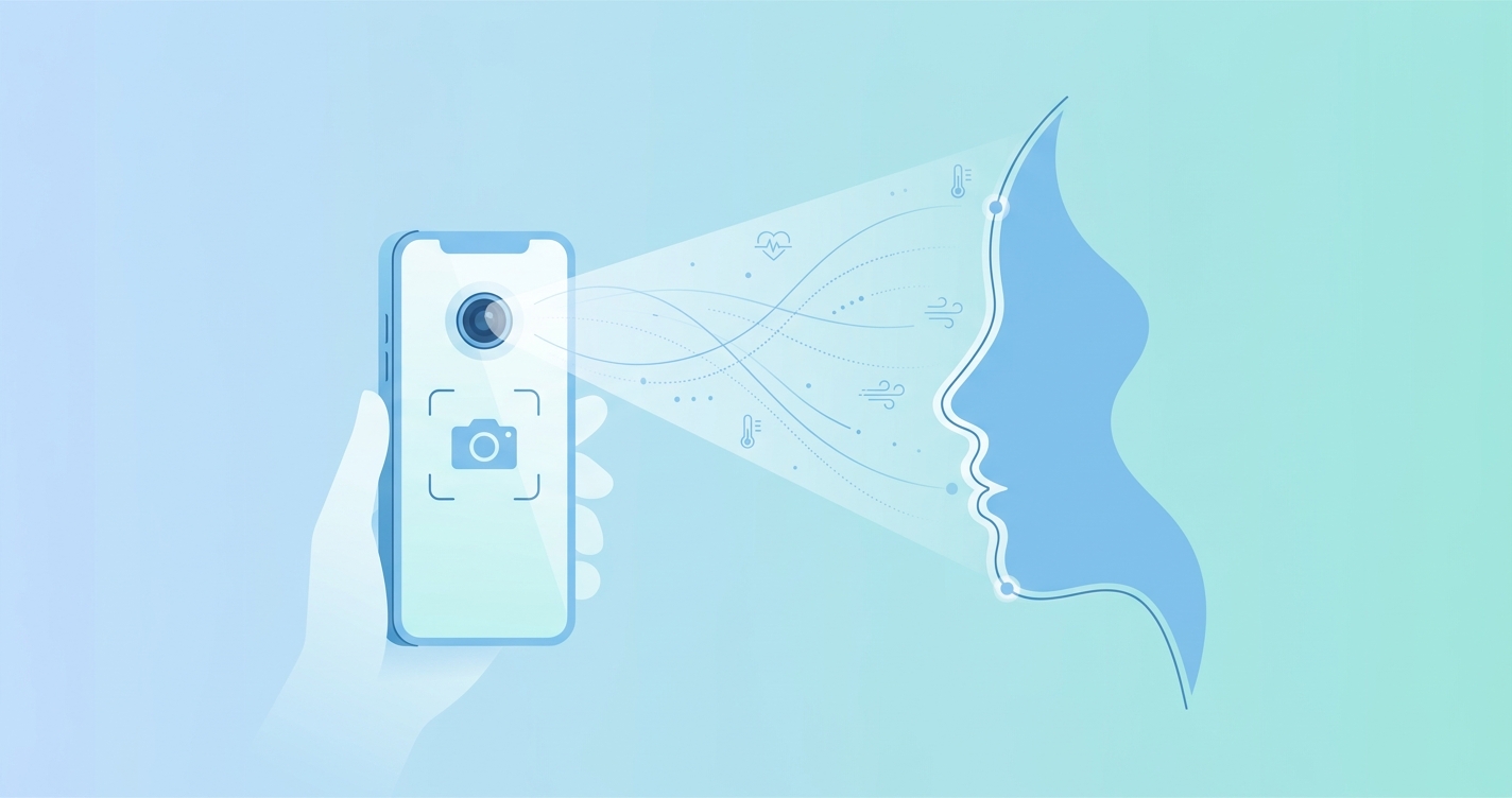
You hold your phone up, look at the camera for half a minute, and get a readout of your heart rate, breathing rate, and stress level. It feels like magic, or maybe like nonsense. But what actually happens during a 30-second face scan that turns a selfie camera into a health sensor? The answer involves blood, light, and some surprisingly clever math.
"Remote photoplethysmography extracts cardiovascular signals from ordinary video of the human face by detecting subtle changes in skin color caused by pulsatile blood flow." — Verkruysse, Svaasand & Nelson, Optics Express, 2008
The first two seconds: finding your face
The moment you open a camera-based vitals app and position yourself in front of the lens, the software runs face detection to locate your head in the video frame. This is the same type of facial landmark detection used across computer vision, identifying the boundaries of your forehead, cheeks, and nose.
What happens next is less obvious. The system picks specific regions of interest (ROIs) on your face where blood perfusion is strongest. Researchers at MIT found that the forehead and cheeks produce the cleanest cardiovascular signals because the skin there is relatively thin and well-vascularized (Poh, McDuff & Picard, Optics Express, 2010). The software locks onto these areas and begins tracking them frame by frame.
If you move your head slightly, motion compensation algorithms adjust the ROI boundaries in real time. Early rPPG systems fell apart with even minor head movement, but modern implementations handle normal fidgeting without losing the signal.
Seconds 3 through 10: reading color shifts you can't see
Here's where it gets interesting. Every time your heart beats, a small wave of blood pushes through the capillaries beneath your facial skin. That blood absorbs and reflects light differently depending on its volume. Your skin color changes with each heartbeat — not enough for your eyes to notice, but enough for a CMOS camera sensor to pick up.
The software separates each video frame into red, green, and blue color channels and tracks intensity changes across them. The green channel carries the strongest signal because hemoglobin absorbs green light more than red or blue. This was first demonstrated by Verkruysse and colleagues in 2008 and has been confirmed repeatedly since then, including by Wang et al. in their 2017 study published in IEEE Transactions on Biomedical Engineering where they introduced the Plane-Orthogonal-to-Skin (POS) algorithm.
At this point, the raw data looks like noise. Ambient lighting fluctuations, tiny camera vibrations, and involuntary facial micro-movements all create interference. The actual pulse signal is buried under several layers of artifacts.
Seconds 10 through 20: separating signal from noise
Signal processing is where rPPG systems earn their keep. The raw pixel intensity data goes through a pipeline that typically includes:
- Bandpass filtering to isolate frequencies in the physiological range (roughly 0.7 to 4 Hz for heart rate, which covers 42 to 240 BPM)
- Independent Component Analysis (ICA) or similar blind source separation techniques to untangle the pulse signal from motion artifacts
- Adaptive noise cancellation that accounts for ambient lighting changes during the scan
A 2024 study by Di Lernia et al. in Behavior Research Methods showed that modern rPPG algorithms can extract reliable heart rate measurements from ordinary webcam footage recorded in uncontrolled environments — home offices, living rooms, variable lighting. The gap between laboratory performance and real-world performance has narrowed considerably over the past five years.
During this phase, the system is also accumulating enough cardiac cycles to produce a reliable measurement. At a resting heart rate of 70 BPM, you'll complete roughly 12 heartbeats in 10 seconds. More cycles mean better averaging and higher confidence in the final number.
What each color channel contributes
| Color channel | Primary role | Why it matters |
|---|---|---|
| Green | Strongest pulse signal | Hemoglobin absorbs green light most effectively |
| Red | Secondary pulse data | Useful for SpO2 estimation via ratio analysis |
| Blue | Reference and noise | Helps isolate motion artifacts from blood volume changes |
| Combined (RGB) | Multi-channel fusion | Algorithms like POS and CHROM use ratios between channels to cancel noise |
Seconds 20 through 28: computing your vitals
With a clean pulse waveform extracted, the system calculates your vital signs. Heart rate is the most straightforward — count the peaks in the waveform and convert to beats per minute.
Respiratory rate comes from a different signal characteristic. Breathing causes cyclical changes in blood pressure and venous return, which modulate the amplitude and baseline of the pulse waveform. By analyzing these slower oscillations (typically 0.15 to 0.4 Hz, corresponding to 9 to 24 breaths per minute), the system extracts your breathing rate from the same video data.
Heart rate variability (HRV) requires measuring the time intervals between successive heartbeats with enough precision to detect beat-to-beat variation. This is harder from video than from an ECG, but a 2025 review published in Frontiers in Digital Health by multiple research groups confirmed that short-term HRV metrics from rPPG are reaching practical reliability for wellness screening, if not yet clinical-grade diagnostics.
Blood oxygen estimation (SpO2) follows a different path entirely. Traditional pulse oximetry compares absorption of red versus infrared light through tissue. Camera-based approaches approximate this by analyzing the ratio of red to blue channel pulsatile components. The physics are similar but the signal-to-noise ratio is lower, which is why camera SpO2 readings carry wider confidence intervals than those from a finger clip.
What a 30-second scan measures vs. traditional methods
| Vital sign | 30-second face scan | Traditional method | Scan approach |
|---|---|---|---|
| Heart rate | Peak detection in pulse waveform | ECG electrodes or pulse oximeter clip | Green channel frequency analysis |
| Respiratory rate | Amplitude modulation of pulse signal | Chest strap or manual counting | Low-frequency envelope extraction |
| Heart rate variability | Beat-to-beat interval analysis | Clinical ECG with electrode placement | Inter-peak timing from video waveform |
| Blood oxygen (SpO2) | Red/blue channel ratio analysis | Finger pulse oximeter with IR LED | Optical absorption ratio approximation |
| Stress index | HRV-derived autonomic balance | Clinical HRV assessment | Sympathetic/parasympathetic ratio from beat intervals |
The final two seconds: confidence scoring
The scan doesn't just spit out numbers. Good implementations run confidence checks on the output. If too much motion occurred during recording, or if lighting was poor, the system flags the measurement as low-confidence or asks you to try again.
A December 2025 study from Bielefeld University, published in npj Digital Medicine by Acharya et al., found that rPPG accuracy drops at elevated heart rates and under low illumination. That finding matters for design: a well-built app needs to know when its readings are trustworthy and when they aren't, and communicate that honestly.
This is also where the 30-second measurement window earns its place. Shorter scans (10 to 15 seconds) can produce a heart rate estimate, but they capture fewer cardiac cycles and leave less room for noise averaging. Thirty seconds hits a practical sweet spot: long enough for reliable multi-vital measurement, short enough that people actually finish the scan.
Why the face and not your wrist or finger
You might wonder why these systems point at your face rather than, say, your hand. Three reasons.
The face has dense superficial vasculature — blood vessels sit close to the surface across a large area. The forehead alone provides a stable, relatively flat measurement surface that stays within the camera frame naturally. And unlike fingers or wrists, your face is already centered in the camera view during a typical phone interaction.
Researchers have experimented with palm-based and wrist-based rPPG, but the face consistently produces stronger signals with lower noise. The ROI is larger, the skin is thinner, and the geometry between camera and skin surface is more consistent.
What affects your scan quality
Not all 30-second scans are equal. Several factors influence measurement quality:
- Lighting: natural daylight or steady indoor lighting works best. Flickering fluorescent lights can introduce 50/60 Hz interference that overlaps with physiological frequencies.
- Skin tone: higher melanin concentration absorbs more light across all channels, reducing signal amplitude. Modern algorithms account for this, but the signal-to-noise ratio is inherently lower for darker skin tones. The research community is actively working on this gap.
- Motion: holding reasonably still matters. Current systems handle mild head movement, but talking, chewing, or large gestures degrade the signal.
- Camera quality: higher resolution and higher frame rate cameras produce cleaner data. Most modern smartphones (30 fps, 1080p or higher) exceed the minimum requirements.
Current research and where it's heading
The field is moving fast. A comprehensive 2025 review in Frontiers in Digital Health surveyed hundreds of rPPG studies and found that heart rate and respiratory rate measurement have reached practical maturity, while blood pressure estimation and more advanced health indicators are still in earlier stages of validation.
Deep learning has changed the game. Rather than hand-crafting signal processing pipelines, researchers are training neural networks end-to-end on video-to-vitals tasks. A November 2025 paper by Taixi Chen and Yiu-ming Cheung on arXiv introduced TYrPPG, a simplified model architecture that improved heart rate estimation accuracy while reducing computational requirements — important for running these algorithms on a phone rather than a server.
The 30-second face scan you take today is meaningfully better than what was possible two years ago, and the version available in another two years will likely handle conditions that currently cause measurement failures.
Frequently asked questions
Is a 30-second face scan accurate?
For heart rate, modern rPPG algorithms produce results within a few BPM of clinical-grade devices under good conditions. Respiratory rate is somewhat less precise. Accuracy depends on lighting, how still you hold, and the specific algorithms used. These readings are useful for wellness tracking and trend monitoring, though they aren't replacements for clinical measurement.
Does the camera actually see my blood?
Not directly. The camera detects changes in how your skin reflects light, which correlate with blood volume changes beneath the surface. It's measuring an optical effect of blood flow, not imaging the blood itself.
Can it work in the dark?
Poorly or not at all. rPPG needs enough ambient light for the camera to detect subtle color changes. Very low lighting reduces signal quality to the point where measurements become unreliable. The Acharya et al. (2025) study in npj Digital Medicine specifically documented this limitation.
Why 30 seconds and not 10 or 60?
Thirty seconds captures enough heartbeat cycles (roughly 30 to 40 at rest) for reliable averaging while remaining short enough for practical daily use. Shorter scans sacrifice reliability. Longer scans improve accuracy marginally but test user patience.
Where this goes from here
Camera-based health sensing is one of those technologies that sounds implausible until you understand the physics. Light interacts with blood. Cameras detect light. Algorithms extract the signal. Thirty seconds later, you know your heart rate.
Companies like Circadify are building on this science to make contactless vital sign measurement accessible to anyone with a smartphone. The scan itself is straightforward. The decade of research behind it is anything but.
If you're curious about the broader technology behind these scans, our piece on what rPPG is and how your phone reads vital signs goes deeper into the underlying science, and our comparison of rPPG and pulse oximeters puts camera-based measurement in context against traditional devices.